
Status: manuscript submitted for peer review (October 2026). These are preliminary, unpublished findings from a retrospective, uncontrolled study. They have not yet been independently reviewed and may change after peer review.
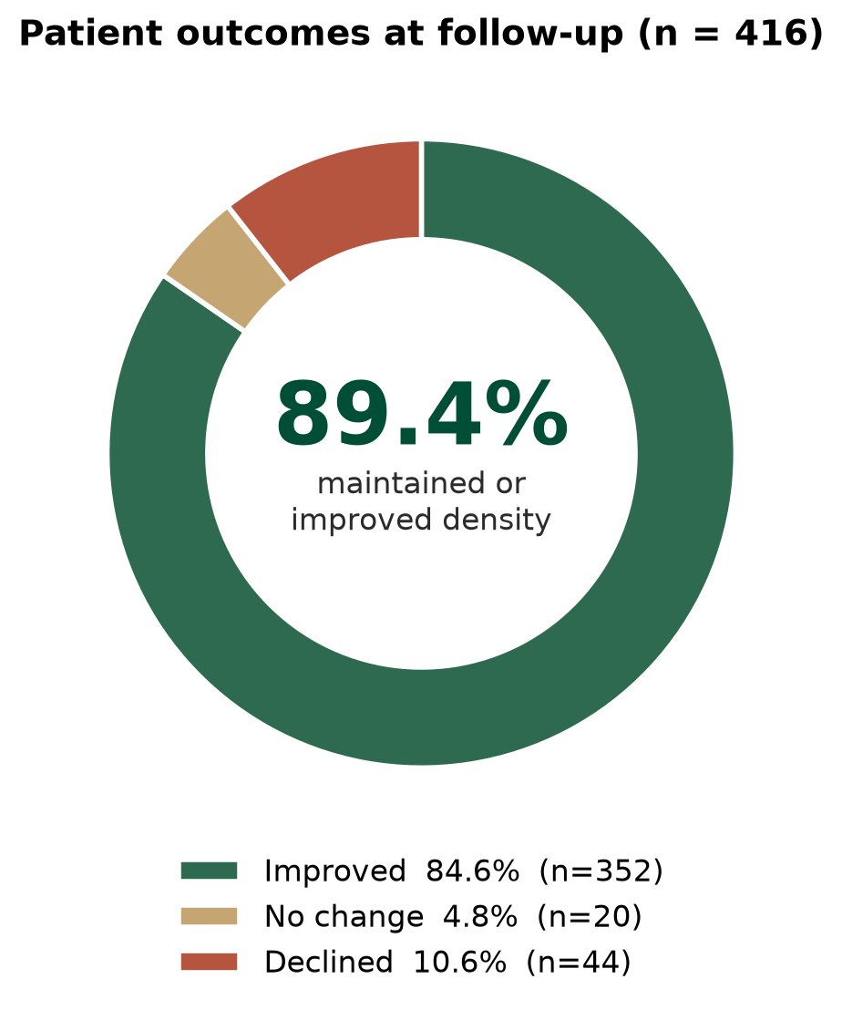
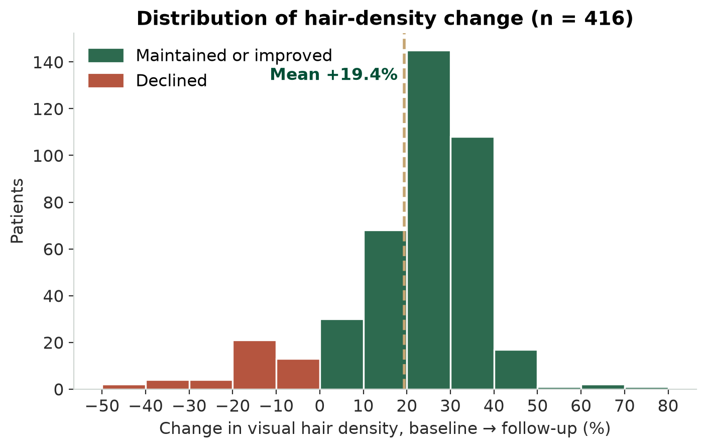
In an internal review of 416 hair-restoration patients photographed at baseline and again a median of 366 days later, 89.4% showed hair density that was the same or better than their starting point. 84.6% showed a visible increase. Mean change in visual density was +19.4% (95% CI 17.8–20.9).
“Visual hair density” is a percentage change estimated from standardized clinical photographs — it is not a follicle count. Individual results vary; some patients saw no change and some lost density. See the full breakdown and limitations below.
Single-session result: among the 190 patients who had one course of therapy with no retreatment, 88.9% maintained or improved density — essentially the same as the full cohort.
Key numbers
The data
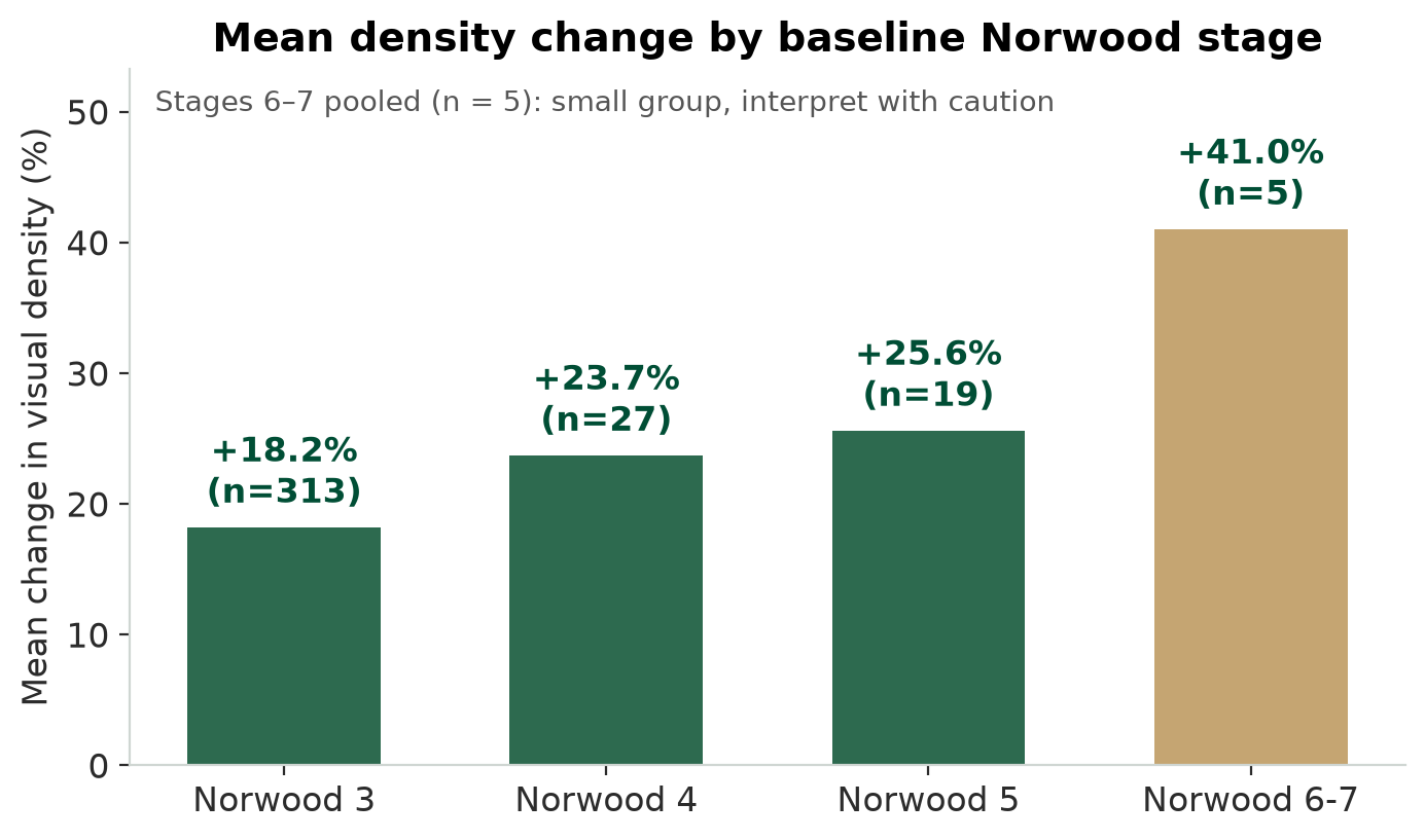
| Baseline stage | Patients | Mean density change | Maintained or improved |
|---|---|---|---|
| Norwood 3 | 313 | +18.2% | 87.2% |
| Norwood 4 | 27 | +23.7% | 96.3% |
| Norwood 5 | 19 | +25.6% | 94.7% |
| Norwood 6–7 | 5 | +41.0% | 100.0% |
| All patients | 416 | +19.4% | 89.4% |
Single session vs. retreatment pathway
| Group | Patients | Mean density change | Visibly improved | Maintained or improved |
|---|---|---|---|---|
| Single session | 190 | +18.6% | 84.2% | 88.9% |
| Retreatment pathway | 226 | +20.0% | 85.0% | 89.8% |
“Single session” = one course of therapy with no retreatment procedure on record. Patients on the retreatment pathway had a second course during the photographic interval. The two groups responded almost identically, so the headline figure reflects a single session.
How the study was done
Who was included
Of 2,112 hair-restoration patients with archived clinical photographs across our 5 U.S. clinics, 642 had photographs on two or more dates at least 60 days apart. After excluding pairs where the photographs could not be fairly compared (different angle, lighting, hair length, or immediate post-procedure images), 416 patients remained. Median time between photographs was 366 days.
What was measured
Baseline and follow-up photographs were scored for percent change in visual hair density under a fixed rubric by an AI vision rater with pre-specified confidence gating — pairs the rater could not score confidently were excluded rather than guessed. Baseline Norwood stage was assigned from the baseline photographs.
What this study is not
- Not a controlled trial. There is no untreated comparison group, so these numbers describe what happened to treated patients — they cannot prove the therapy caused the change.
- Not a follicle count. Visual density from photographs is an estimate, not a trichoscopic measurement.
- Not every patient. Only patients who returned for follow-up photographs are included. Patients who did not return may have had different outcomes.
- Not final. The manuscript is under peer review. Findings may be revised.
Important information
Preliminary and unpublished. These results come from an internal retrospective study conducted by Springs Rejuvenation. The manuscript has been submitted to a peer-reviewed journal and has not yet been accepted or published. Results are preliminary and may change following peer review.
Not FDA approved. Stem cell and exosome therapies are not FDA approved and are considered investigational. These statements have not been evaluated by the Food and Drug Administration and are not intended to diagnose, treat, cure, or prevent any disease.
Individual results vary. The figures on this page are group averages from one clinic network's patients. They are not a prediction or guarantee of any individual's outcome. Some patients in this study saw no change, and some lost density.
Study design. Retrospective, uncontrolled, single-network cohort using AI-assisted photographic grading. It cannot establish causation and is subject to selection bias, since only patients who returned for follow-up photography are represented.
Conflict of interest. This study was conducted and funded by Springs Rejuvenation, which provides the therapy evaluated. The authors are affiliated with Springs Rejuvenation.
Not medical advice. This page is for educational purposes only. It does not replace a consultation with a licensed physician who can evaluate your individual situation.
Reviewed for medical accuracy by Dr. Charles Pereyra, MD, Medical Director, Springs Rejuvenation. Data frozen October 15, 2026.